
A subject of wide interest in modern Biophysics, the Life and Material Sciences is the noninvasive characterization of the structure and dynamics of molecular species ranging from a single molecule to a complex heterogeneous system, i.e. a living cell or tissues, through advanced optical microscopy methods. Two main areas that represent different aspects of our research activities in this field encompass Coherent Raman Scattering (CRS) Microscopy and Single-Molecule Fluorescence Biophysics:
Research Interests
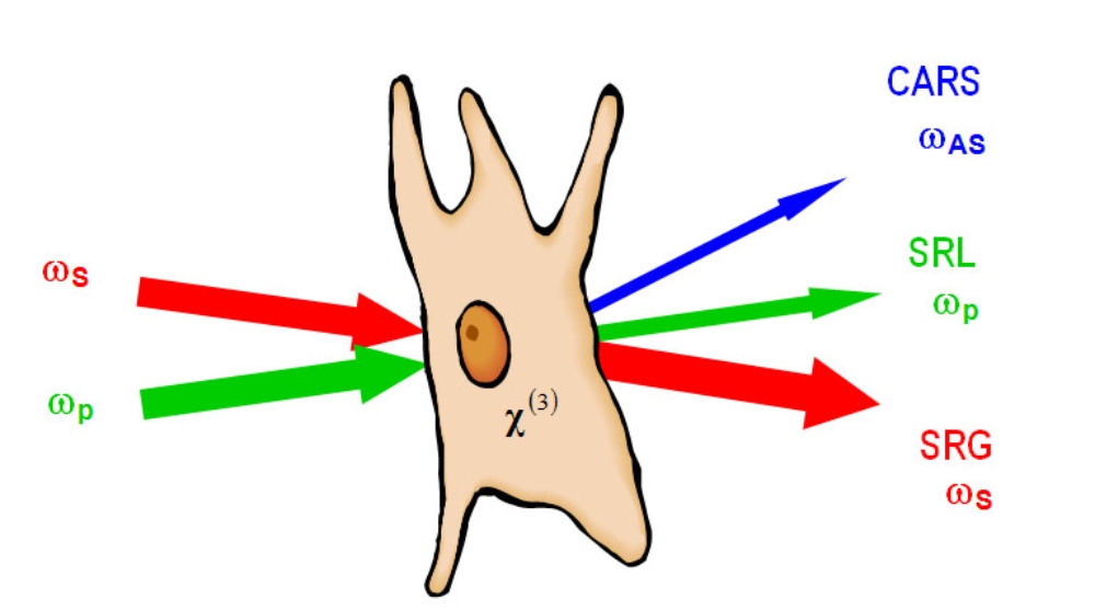
A subject of wide interest in the modern Material and Life Sciences is the noninvasive characterization of the structure and dynamics of molecular species within a complex heterogeneous system, i.e. a living cell or tissues, through advanced optical microscopy. For molecular species that either do not fluoresce or cannot tolerate the toxicity associated with fluorophore labeling, their intrinsic molecular vibrational properties can be used as contrast mechanisms in vibrational microscopy. The signal-to-background limitations inherent to conventional spontaneous Raman and IR detection schemes can be circumvented by exploiting the coherent driving and detection of Raman modes in coherent anti-Stokes Raman scattering (CARS) and stimulated Raman scattering (SRS) microscopy, the latter of which was first successfully demonstrated by Nandakumar et al. in 2005 in Stuttgart. CRS detection significantly enhances the Raman scattering signal by several orders of magnitude. Our current research activities are focusing on the methodological advancement of CRS microscopy for the quantitative and three-dimensional mapping of chemical and physical structure information of biomolecular species under in-vivo conditions.
For recent Review articles see:
- A. Volkmer (2005) J. Phys. D: Appl. Phys. 38:R59-R81 (Topical Review).
- A. Volkmer, Chapter 6: Coherent Raman scattering microscopy, in Emerging Biomedical and Pharmaceutical Applications of Raman Spectroscopy Eds. P. Matousek and M. Morris, 2009 (Springer- Verlag, Berlin, Heidelberg, New York, Tokyo, ISBN: 978-3-642-02648-5).
Single-Molecule Fluorescence Microscopy, using biochemical techniques to obtain chemical selectivity, has developed into a very powerful tool in biophysical and biomedical research that provides the ultimate level of sensitivity and avoids ensemble averaging. Our group is focusing on the further development and application of advanced single-molecule spectroscopy techniques that are instrumental in the study of fundamental problems in Biophysics. Current research activities include:
- the analysis and control of conformational dynamics in a membrane transporter protein “nano-machine”,
- the probing of hydrodynamic properties and dynamics of proteins by a novel rotational fluorescence correlation spectroscopy,
- and the study of the photon statistics in the fluorescence from single pigment-protein complexes.
Members of the Group
- Ph.D. Student
Stefan Gomes da Costa,Dipl. phys.
Tel: 685 66685
E-Mail - Ph.D. Student
Wan Hui, M.Sc.
Tel: 685 66685
E-Mail - Principal Investigator
Andreas Volkmer, Ph.D.
Tel: 685 65236
E-Mail